

FLI provides starting grants to projects involving at least two distant laboratories in France to maintain the scientific expertise of the teams operating the imaging platforms at the highest level. The four scientific expertise networks, RE1 in the field of imaging agents, RE2 in the field of instrumentation and technological innovation, RE4 in the field of multimodal image analysis carried out this scientific animation program.
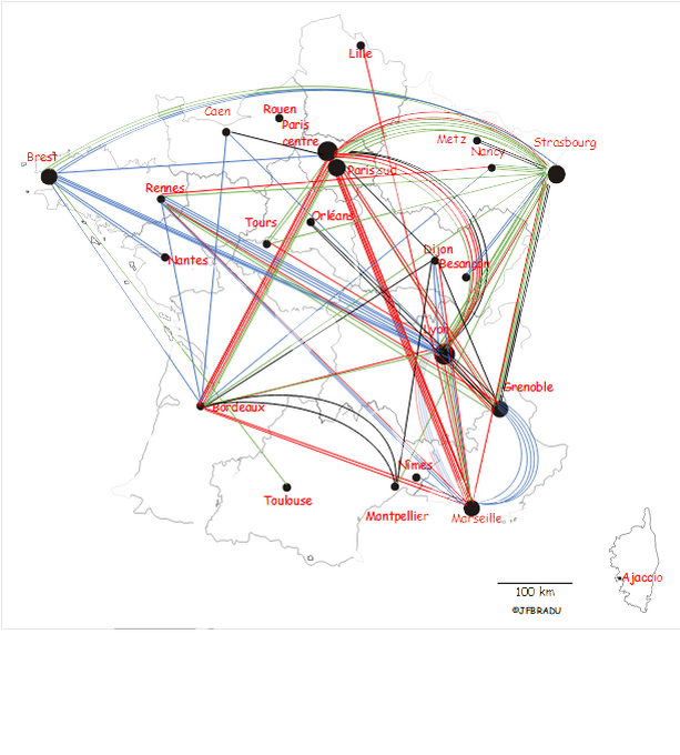
Financial support is granted based on calls for projects launched regularly since 2013 and after selection by the steering committee of the four networks of expertise. The calls for projects are open to all “FLI” and “non-FLI” laboratories. Since 2013, 141 projects of exchange of expertise between at least 2 distant laboratories have been funded for a total amount of approximately 2.6 M€.
These exchanges create a dense network of laboratories (see the map below)
Several FLI-supported projects have been at the origin of more ambitious projects funded by national or international funding agencies, demonstrating the leverage effect of FLI’s expertise exchanges.
Since 2013, fifteen projects out of the 141 supported by FLI, have enabled laboratories to raise a total of more than €12M in funding, either from the French Research Agency, foundations, or from European H2020 programs. Examples of projects that originally received support from FLI include:
- IMOP (Interoperative Multimodal Optical Probe) supported by the Cancer Plan,
- ARMONI (single-pixel imaging methods), a JCJC project of the ANR,
- OptimiX (Optimization of the radiation dose for X-ray guided procedures) supported by the ANR PRC,
- ROBOT (Robotics and optical coherence tomography for optical biopsy in the digestive tract),
- M-CUBE, FET-OPEN,
- QuantSURG, ERC,
- MRgHIFU (MRI-guided high-intensity focused ultrasound) supported by the ANR,
- SURGEONAID (Transforming brain surgery by advancing function-guided neuronavigation imaging) funded by Europe EIC Pathfinder,
- NIFUS (Non-invasive focused ultrasound surgery of breast adenocarcinomas) supported by INCa.
In another field, one of the expertise exchanges granted by FLI in 2020, related to the performance evaluation of new sensors for MEG, contributed to the creation of Mag4Health – a startup supported by the CEA – which will market the tested sensors.
The following pages present the expertise exchange projects supported network by network.
Network of Expertise 1: Molecular Imaging Agents (NE1)

Since 2015, the Expertise Network 1, RE1, has financially supported 26 projects for expertise exchanges between research laboratories and imaging platforms in France for a total of €675k. More than 30 different laboratories have benefited from these starting grants. The table below presents the funded projects.
| Year of award | Short title | Project title | Teams / laboratories involved |
| 2015 | PoC | First in man PET study of a radiotracer for Alzheimer’s disease early imaging | CERMEP Lyon |
| 2017 | GRAMM | Imagerie bactérienne par marquage métabolique du lipopolysaccharide | Sondes moléculaires / SHFJ Orsay & Institut Chimie Moléculaire et des Matériaux d’Orsay |
| 2017 | PINK | PET Imaging with Neurotensin analogues in breast cancer | UMR5247 / Montpellier & UMR5287 / Bordeaux |
| 2017 | RADIOPLATIN | radiothérapie ciblée : vectorisation du carboplatine 195m pour le traitement du cancer de l’ovaire | UMR 5247 & U 1194 / Montpellier & NRG Petten Netherland |
| 2017 | FluoNTep | Sonde bimodale analogue de la neuro tensine pour l’imagerie TEP et la chirurgie du cancer du pancréas guidé par FLUOrescence | UMR CNRS 6302 Univ Bourgogne & UMS 28 Université Paris6 (Sorbonne Univ) |
| 2017 | MEDI-SURF | Medical Devices Imaging through surface Modification | UMR 5247 & UMR 5253 & UMR 5221 / Université Montpellier |
| 2017 | ZiFiZiCA | In vivo Zinc detection by MRI using peptidic contrast agents based on the zinc finger sequence and containing Gd3+ chelate | Centre de Biophysique Moléculaire, Orléans & Institut de Recherche Interdisciplinaire de Grenoble / Physico-chimie des Métaux en Biologie, Grenoble |
| 2018 | NIRPER | NIR-laser-switched smart nanocomposites as contrast agents for photoacoustic imaging and photothermally-activated durg-delivery systems for detection and therapy of peritoneal carcinomatosis. | MSC (UMR7057)/ Univ. Paris Diderot Paris & LCBPT Univ. Paris Descartes Paris & LIB (UMR7371) Sorbonne Univ. Paris |
| 2018 | CocTEL | Imagerie de la coqueluche (B. pertussis) par immunoTEP-IRM d’un anticorps spécifique du LOS-A marqué au zirconium-89 | SHFJ Orsay & IDMIT Fontenay-aux-Roses |
| 2018 | GlioSpy | Nouvelles sondes pour l’imagerie photo-accoustique de l’inflammation péri-tumorale et péri-lésionnelle anant et après résection des glioblastomes: information manquante pour la prédiction de la récidive précose chez les patients. | UMR5182, ENS, Lyon & U1039, Grenoble |
| 2018 | Théra-BODIPY | Elaboration de systémes théranostiques à base de BODIPY pour l’imagerie optique et la radiothérapie par activation du bore par les neutrons (BNCT). | IAB U1209 UMR5309 & ICMUB UMR6302 & CERMAV, Grenoble |
| 2018 | BIFPETBR | Double imagerie (fluorescence et TEP) des tumeurs cérébrales gliales par détection des récepteurs aux endothélines. | ICMUB-UMR6302, Dijon & SPI-LEMM, CEA, Saclay & INM (UMR 1051), Montpellier |
| 2021 | BIVALIMAG | Traceurs peptidiques bivalents pour le ciblage tumoral en imagerie nucléaire | UMS28 Université Paris6 & INCIA CNRS UMR5287 Bordeaux & ICMUB UMR6302 |
| 2021 | FLUOTENSINE | Late and selective fluorination of neurotensin probes for improved pre-therapeutic PET imaging | INCIA CNRS UMR5287 Bordeaux & IBMM Montpellier |
| 2021 | SEP’N’TEP | Nouveaux radio traceurs et biomarqueurs de l’inflammation en Tomographie par Emission de Positons (TEP) dans la sclérose en plaques (SEP) | BioMaps SHFJ CEA Orsay & BIORAN-CERMEP Lyon |
| 2021 | NANOLIGATION | Développement et évaluation d’une méthode de conjugaison enzymatique site-spécifique de nanoparticules sur des anticorps anti-PD-L1 pour des applications en théranostique | BioMaps SHFJ CEA Orsay & ILM/FENNEC Lyon |
| 2021 | I2HAVC | Approche multimodale par imagerie TEP/IRM pour la détection de l’hypoxie et la compréhension de la relation hypoxie-inflammation dans les AVC : évaluation de nouveaux radio pharmaceutiques 18F-fluorosulfonitroimidazoliques | LDM/TEP Caen & PhIND |
| 2021 | ImaFibro | Validation de la Lysyl oxidase-like 2 (LOXL2) et du Fibroblast Activation Protein (FAP) comme cibles d’imagerie pour la quantification et le suivi de la réponse thérapeutique dans un modèle murin de fibrose pulmonaire | ICMUB UMR6302 Dijon & CLCC Georges-François Leclerc Dijon |
| 2022 | COFAKTEUR | Développement de radiotraceurs covalents de la protéine FAK comme compagnon thérapeutique pour l’imagerie TEP en oncologie |
BIOMAPS SHFJ Orsay & UNIV PARIS V LCBPT Paris |
| 2022 | ECM-TRACER | Développement d’un radiopharmaceutique peptidique ciblant la Tenascin-C pour l’imagerie TEP de la matrice extracellulaire de l’adénocarcinome pancréatique | INSERM U1194 MONTPELLIER & UMR CNRS 6302 |
| 2022 | HYPER TEP | Ligands du récepteur 5-HT4 fluorés pour le diagnostic de l’hyperaldostéronisme en TEP | CERMN Caen & BIOMAPS SHFJ Orsay |
| 2022 | PULMOTEP | Imagerie des récepteurs NMDA pulmonaires : vers un radiotraceur innovant pour l’imagerie du remodelage vasculaire pulmonaire en TEP | BIOMAPS SHFJ Orsay & UMR S 999 Le Plessis-Robinson & BIOCIS Paris Saclay |
| 2022 | SMILE | Agents de contrastes émettant dans le SWIR pour l’imagerie in vivo à hautes résolutions des cancers | IAB GRENOBLE & IPHC STRASBOURG & DCM GRENOBLE |
| 2022 | IMPACTION | Nouvelles sondes pour l’IMagerie PhotoACousTique de l’InflammatiON | CAMB UMR 7199 Srasbourg & TIMC UMR 5525 Grenoble & OPTIMA LIPHY & MOVE LIPHY Grenoble |
| 2022 | PSICADELIC | Ph sensitive MRI contrast agent based on nitrone for the detection of triple negative breastcancer | DCM Grenoble & CMB Orléans |
Network of expertise 2: Instrumentation & Innovations Technologiques (NE2)

Since 2013, FLI’s Network of Expertise 2, NE2, has provided 39 starting grants to promote expertise exchange projects, including 16 since 2021, for a total of 629k€. The average grant amount is ~21 k€. These financial supports have been awarded to 31 different laboratories. Of the ongoing projects, every exchange, except one, involves at least 2 hubs. Four (4) exchanges involve 3 hubs and 1 exchange involves 4 hubs. Finally, 2 projects at the interface with NE1 and NE4 have received financial support from each of the networks. Thirteen (13) projects are in the field of MRI, 2 in US, 1 in MEG, 1 in PET, 1 in acousto-optics and 1 mixed US-MR project. Four (4) projects focus on protocol robustness and safety issues in ultra-high field MRI, 6 on the development of MRI biomarkers (MS, lipid content, brain oxygenation, heart transplant viability, high resolution multiparametric imaging, free surface fMRI), 3 on miniature MRI coils intracardiac- deployable or implantable in the brain (non-exhaustive list). Five projects propose new imaging methods or disruptive concepts, in-vivo acousto-optics, wireless implantable US transducer array, anisotropic/non-linear elastography by MR or US, MEG with laser pumped magnetometers.
The table below summarizes the expertise exchange projects supported since 2013.
| Year of award | Short title | Title of the project | Teams / laboratories involved |
| 2013 | MR Spectroscopy | Hyperpolarisation et microsonde RMN pour la spectroscopie in vivo du 13C | Bordeaux & Paris Sud |
| 2013 | UHF MRI | Développement de capteurs multiéléments pour l’imagerie par RM parallèle du petit animal à ultra haut champs magnétique. | CREATIS Lyon & NeuroSpin Saclay |
| 2013 | SPICO-imaging | Imagerie de la moelle épinière et développements d’outils pour l’analyse des données | Marseille & Lyon |
| 2013 | qSMR | Imagerie spectroscopique du petit animal : quantification, visualisation, standardisation | CREATIS Lyon & U1216 GIN Grenoble |
| 2013 | MR spectroscopy at UHF | Outils méthodologiques pour la spectroscopie RM à haut Champ | NeuroSpin Saclay & CRMBM Marseille |
| 2014 | Optimi DTI | Optimisation DTI sur rongeur | U1216 GIN Grenoble & NeuroSpin Saclay |
| 2014 | TofNIRS | Structuration nationale des forces en imagerie non invasive du fonctionnement et des pathologies cérébrales par spectroscopie proche infrarouge résolue en temps de vol | Lyon &Strasbourg &Grenoble |
| 2014 | EndoscopieNon Lin | Imagerie du vivant par endoscopie non-linéaire | MOSAIC Institut Fresnel Marseille & Lille |
| 2014 | 13PhSMR | Mise au point, validation et application d’une séquence d’imagerie spectroscopique du phosphore 31 à 3T pour l’étude métabolique du muscle squelettique à l’exercice | CRMBM Marseille & Dijon |
| 2014 | META7 | Shimming Radio Fréquence Passif pour IRM 7 T | NeuroSpin Saclay & Institut Frenesl Marseille |
| 2015 | US-Contrast | Nouvelles méthodologie en imagerie de contraste ultrasonore : modélisation de l’écoulement des agents de contraste ultrasonores dans une tumeur | Paris Sud /Strasbourg |
| 2015 | IRM Quantitative Haut et Très Haut Champs (qMRI7) | méthodes d’IRM quantitatives multiparamétriques à haut et très haut champ (3T et 7T) | NeuroSpin Saclay & ICUBE Strasbourg |
| 2015 | Seno-IRM | Antenne dédiée à l’IRM du sein | IADI Nancy & IR4M Paris Sud |
| 2015 | qMRI | Missions pour le montage d’un projet collaboratif (type ANR) pour « IRM quantitative » | IR4M Orsay & CRMBM Marseille & ICUBE Strasbourg & ICM-CENIR Paris |
| 2017 | ENDOMouse | Endoscopie Multimodale et IRM endoluminale dans un modèle murin d’inflammation colorectale | Creatis Lyon & CHRU Tours |
| 2017 | MAPSSIC | Développement d’une sonde télémétrique CMOS pour la mesure de radiophamaceutiques chez l’animal éveillé et libre de ses mouvements | IMNC/UMR 8165 Orsay & CPPM UMR 7346 Marseille & IPHC UMR 7178 Strasbourg |
| 2017 | REMI | Réseau d’entraite pour les études multicentriques en IRM | ICM Paris & VISAGES Rennes & IRMaGe GIN Grenoble & CIC-IT Nancy & INT Marseille |
| 2018 | RFI 7T | Réseau français des utilisateurs d’IRM 7T | NeuroSpin Saclay & CRMBM Marseille |
| 2018 | MRI TSAFETY | Sécurité thermique dans le contexte de l’IRM à ultra haut champ magnétique | Lyric Bordeaux & Neurospin Saclay |
| 2018 | AEC MRI | Développement d’antennes endoscopiques pour l’IRM cardiaque | Lyric Bordeaux & IR4M Orsay |
| 2017 | Simultaneous MRI | Simultaneous PET-MR dynamic acquisitions for measuring perfusion, vascular permeability, and inflammation in stoke | CERMEP Lyon & CREATIS Lyon & U1216 GIN Grenoble |
| 2018 | FFC-IR4M / Relaxométrie RMN | Relaxométrie RMN – Mise en place et validation multi-centrique multi constructeur à 1.5, 3 et 7T | CRMBM UMR 7339 Marseille & IR4M UMR 8081 Orsay |
| 2019 | Micro NMR | Micro-sondes RMN à très hauts champs | Neurospin Saclay & UMR 5255 Bordeaux |
| 2020 | MEG-PMLG | les Apports des magnétomètres à pompage optique pour la détection des activités électromagnétiques en MEG | CEA LETI Grenoble & ICM Paris & CNRS UMR 7225 UMR 1106/CERMEP Lyon |
| 2022 | CRYOXYbrain | Développements et évaluation de méthodes pour l’imagerie de l’oxygénation cérébrale | MIRCEN Fontenay-aux-Roses & GIN Grenoble |
| 2022 | QUALIPSI | Quantification lipidique par EPSI –« QUALIPSI » | CREATIS Lyon & CRMBM Marseille & BioMaps Orsay |
| 2022 | UHR PET | Conception d’une chaise de contention pour PNH éveillé, adaptée au prototype d’imageur TEP haute résolution dédié cerveau (UHR PET) | CERMEP Lyon & BioMaps SHFJ Orsay |
| 2022 | MALUS | Quantification des propriétés Mécaniques, Anisotropes et non-Linéaires du muscle en élastographie UltraSonore | ICUBE/BioMaps |
| 2022 | TRAME | Transmission parallèle pour l’IRM 7T de la moelle épinière | NEUROSPIN/CRMBM |
| 2022 | BODESS | Development of an B0 fluctuation-insensitive 3D DESS sequence for human brain T2 mapping at high and ultra-high magnetic fields | CRMSB Bordeaux & CRMBM Marseille |
| 2022 | T1-M3C-SEP | Vers un usage Multicentrique de la technique de relaxométrie T1-MP2RAGE au niveau de la Moelle épinière et du Cerveau – Application à la Sclérose En Plaques | Empenn Rennes & CRMBM CEMEREM Marseille |
| 2022 | RAVIOLE | imageRie Acousto-optique in VIvO pour Le suivi de tumeurs sur pEtit animal | Institut Langevin ESPCI /Laboratoire UTCBS – Université de Paris Cité |
| 2022 | HARMONI | Initiative pour l’harmonisation et la standardisation des protocoles et bonnes pratiques d’acquisition en IRM au sein du Réseau Français des IRM Ultra-Haut Champ 7T | CRMBM Marseille & CHU POITIERS & NEUROSPIN Saclay |
| 2022 | IDEAL | rm endovasculaire cardiaque plastronique | AMPERE CNRS Lyon & IHU LYRIC – RMSB Bordeaux & BIOMAPS SHFJ Orsay |
| 2022 | LilipUS | lecture parallèle d’implants sans fils par imagerie ultrasonore ultra-rapide | U1216 GIN Grenoble & U1032 (LabTAU) Lyon |
| 2022 | PRIMATE | IRM multiparamétrique haute résolution du cerveau de PNH | ICUBE Strasbourg & CERMEP Lyon & ICM Paris |
| 2022 | QUANTUM | QUANtification des propriétés élastiques non linéaires de TUMeurs par élastographie US et IRM. | BIOMAPS SHFJ Orsay & INSERM U1149 CRI Paris |
| 2022 | VIAMY | Etude en IRM et spectroscopie de la viabilité myocardique du greffon cardiaque avant transplantation | LIB SORBONNE UNIVERSITE Paris & CRMBM Marseille |
| 2022 | FreeFace4MRI | Optimisation de protocole en IRMf avec une antenne tête ne couvrant pas le visage. | CERMEP Lyon & CIC-IT CHRU Nancy |
Network of Expertise 3: Image Guided Interventional Radiology (NE3)

The Network of Expertise 3 (NE3) has granted financial support to 42 expertise exchange projects since 2013. The average grant amount is ~13k€. All in all, 552k€ has been awarded to 24 different research laboratories. The laboratories are located within hubs or outside the regional and the transverse IAM hubs.
The table below details the exchanges of expertise and the laboratories involved.
| Year of award | Short title | Titel of the project | Teams / laboratories involved |
| 2014 | REALTIMUS | Real time software for MR guided HIFU | IHU – Lyric Bordeaux & Neurospin Paris Saclay |
| 2014 | MUSTANG-FIR | Minimally-invasive UltraSound Therapy under Augmented Navigation Guidance_ pre-investigations on the treatment planning under Fusion Imaging guidance and Robotic control | LabTau (INSERM U1032) Lyon & ICube (UMR CNRS 7357) Strasbourg |
| 2014 | GET/STRIP | Guidage de l’Exérèse Tumorale par Stratégies Temps Réel Innovantes Per-opératoires | CEA Clinatec Grenoble & Neurospin Paris Saclay |
| 2014 | GUIDANCE | Guidage per-opératoire pour l’exérèse de tUmeurs cérébrale basé sur modélisation bIomécanique patient-spécifique Dirigée par l’imAgerie iNterventionnelle éChographiquE | TIMC (UMR CNRS 5525) + Clinatec Grenoble & ICube (UMR CNRS 7357) Strasbourg |
| 2014 | FLUOFIBRE | Imagerie optique de fluorescence interventionnelle en neurochirurgie pour l’assistance au geste opératoire lors des exérèses des gliomes | CREATIS (UMR CNRS 5220) Lyon & IMNC (UMR CNRS 8165) – Orsay |
| 2014 | Porte Aiguille Robotise | Développement d’un porte-aiguilles robotisé à compliance adaptative basé sur un mécanisme de tenségrité pour la radiologie interventionnelle sous IRM | ICube (UMR CNRS 7357) – Strasbourg & LIRMM (UMR CNRS 5506) – Montpellier |
| 2015 | SONOCIB | Évaluation des microbulles ciblant le récepteur de la transferrine pour l’ouverture transitoire de la barrière hémato-encéphalique par ultrasons guidés par imagerie | NeuroSpin – Paris Saclay & U930 Imagerie et cerveau Tours |
| 2015 | 2PFLUODIAG | Imagerie multimodale qualitative et quantitative de biopsies étagées de glioblastome : construction d’une base de donnée tissulaire | IMNC-UMR CNRS 8165 Orsay & Hopital Sainte Anne Paris |
| 2015 | PhaseBrain | Reconstruction des images de phase pour la thermométrie cérébrale et l’imagerie de susceptibilité magnétique à 3T | IHU-Liryc – Bordeaux Université & Institut Langevin/Paris |
| 2015 | ANNFEET | Aide à la navigation de systèmes flexibles Endolumineux par échographie transabdominale | ICube (UMR CNRS 7357) – Strasbourg & IRISA (UMR6074) Rennes |
| 2015 | ExpoMRI | Simulation de champ et de détection visuelle des Personnes pour la validation d’un capteur Portatif d’Exposition IRM | Icube (UMR CNRS 7357) Strasbourg & IADI (UMR947)Nancy |
| 2015 | RTprocessing | Traitement de l’information optique en temps réel pour l’imagerie interventionnelle | CREATIS (UMR CNRS 5220) Lyon & ICube (UMR7357) Strasbourg |
| 2015 | ThermoMetRE | Compensation des mouvements respiratoires en Elastographie par résonance magnétique (ERM) et thermométrie IRM simultanéees | Icube (UMR CNRS 7357) Strasbourg & IHU Lyrics Bordeaux & Institut de Mathématiques de Bordeaux |
| 2017 | FLUODIAG 2 | Imagerie multimodale qualitative et quantitative de biopsies étagées de glioblastome : construction d’une base de donnée tissulaire | IMNC-UMR CNRS 8165 Orsay & Hopital Sainte Anne Paris |
| 2017 | COSMIC | Robot à tubes COncentriqueS pour la MIcroscopie Confocale | NEUROSPIN Saclay & ICube (UMR CNRS 7357) Strasbourg |
| 2017 | ERM interventionnelle passive | Développement d’une méthode d’elastographie par résonance magnétique passive pour la radiologie interventionnelle | ICube (UMR7357) Strasbourg & LabTAU (U1031) Lyon |
| 2017 | VATSnext | Images et modèles pour le guidage d’intervention par vidéothoracoscopie | LTSI (U1099) Rennes & TIMC-IMAG (UMR5525) Grenoble |
| 2017 | MICA | Méthodes avancées pour l’imagerie du cathétérisme Actif | ISIR Paris & LiSSI/UPEC (Université Paris-Créteil) Créteil |
| 2017 | SUNBEAM | Surgery application of Ultrasound based Nonlinear Biomechanical Measurements | Icube (UMR 7357) Strabsourg & Institut Langevin Paris |
| 2017 | IMBO 1 | Méthodes d’imagerie innovantes pour la caractérisation de tissus et substituts osseux | Icube (UMR7357) Strasbourg /IR4M (UMR8081) Orsay & C2N (UMR9001) Paris Saclay |
| 2018 | ECHOPT | Optimisation temps réel de positionnement d’outils chirurgicaux basés sur l’imagerie peropératoire pour la chirurgie laparoscopique et percutanée) | TIMC-IMAG (UMR5525) Grenoble & Icube (UMR7357) Strasbourg |
| 2018 | TSONOAMI | Ultrasons de basse énergie pour l’imagerie et la thérapie cardioprotectrice : preuve de concept et validation expérimentale par IRM sur le modèle porcin dans le contexte de la protection du myocarde lors de la reperfusion post-infarctus | CREATIS (UMR5220) Lyon & LabTAU (U1032) Lyon |
| 2018 | REED | IMNC (UMR8165) Orsay & LCMCP (UMR7574) & LISE (UMR8235) Paris | |
| 2018 | IMBO 2 | Méthodes d’imagerie innovantes pour la caractérisation de tissus et substituts osseux | Icube (UMR7357) Strasbourg & IR4M (UMR8081) Orsay & C2N (UMR9001) Paris |
| 2018 | HIFUcgMRI | Générateur compact d’ultrasons à haute intensité focalisés, guidé par Imagerie par Résonance Magnétique | C2N (UMR9001) Paris & Icube (UMR7357) Strasbourg |
| 2018 | MUSE-HiFU | Monltoring UltraSonorE des HiFU | LabTAU (U1032) Lyon & LMA (Laboratoire de mécanique et d’accoustique) UMR 7031 Marseille |
| 2018 | COSMIC 2 | Robot à tubes COncentriqueS pour la MIcroscopie Confocale | UMR 6174 (Femto-ST) Besançon & ICube (UMR7357) Strasbourg |
| 2018 | ROBOPTX | Optimisation de la Pose d’un Arceau Robotisé pour Minimiser les Doses Reçues par les cliniciens et le Patient | ICube (UMR7357) Strasbourg & LaTIM (UMR 1101) Brest |
| 2018 | FAMTASTIC | Fusion d’imAges Multimodales pour la Thérapie, l’ASsisTance et le guidage en Insuffisance Cardiaque | LTSI (U1099)+TIMC-IMAG (UMR CNRS 5525) |
| 2018 | SURGEONAID | Real time intraoperative optical imaging of cerebral hemodynamics for neurosurgery guidance | CNRS DR7 (CREATIS) Lyon & ICUBE Strasbourg |
| 2018 | APRAGUI | Apprentissage Profond en radiothérapie guidée par l’image | Icube Strasbourg & CREATIS Lyon & LTSI Rennes & LaTIM Brest |
| 2018 | NC-ELASTO | Fully non-contact optical quantitative elastography using time reversal approach | CNRS DR10 (Icube) Strasbourg & LabTau Lyon |
| 2018 | CURAC | Cartograpie Ultrasonore électromécanique haute Résolution des Arythmies Cardiaques pour guider le traitement local par ablation | LABTAU Lyon & IHU – Lyric Bordeaux |
| 2018 | CAVIMUT | Technique innovante de contrôle de cavitation durant les séquences d’ultrasons thérapeutiques | IR4M Orsay & GREMAN Tours |
| 2021 | DEMAISED | Développement de méthodes d’imagerie innovantes pour la micro vascularisation des gliomes cérébraux | iBrain – Tours & IRIT Toulouse |
| 2021 | M-QUS | Monitoring Quantitatif UltraSonore des traitements par HIFU | LABTAU Lyon & LMA (Laboratoire de mécanique et d’accoustique) UMR 7031Marseille |
| 2021 | SPIM | Spectro-Imagerie optique Multimodalités pour la prise en charge diagnostique des plaies chroniques. | IRCAD Strasbourg & CRAN Nancy |
| 2021 | BONEGHOST | Fantôme osseux à haute-fidélité morphomécanique pour les procédures de cimentoplastie. | ICUBE-AVR Strasbourg & IADI Nancy |
| 2021 | IMCOLL | Imagerie multimodale de structures collagéniques : membrane et mousse. | ICUBE-MMB Strasbourg & LEM3 Metz |
| 2022 | M-QUS | Monitoring Quantitatif UltraSonore des traitements par HIFU | LABTAU Lyon & LMA (Laboratoire de mécanique et d’accoustique) UMR 7031 Marseille |
| 2022 | TENDEMBOL | TendEmbol (Evaluation du retentissement vasculaire après embolisation dans un modèle porcin de tendinopathie – Evaluation de la néoangiogénèse par imagerie isotopique et histologie). | CERIMED Marseille & CHU NIMES |
| 2022 | RADIUS | Registration of biomechanicAI moDels wlth UltraSound images | ICUBE Strasbourg |
| 2022 | ViscOptique | Imagerie optique des propriétés viscoélastiques de tissus biologiques | ICUBE Strasbourg & LABTAU Lyon |
Network of Expertise 4: Multimodal Image Analysis (NE4)

Since the creation of FLI, the network of Expertise 4 (NE4) awarded 44 starting grants “Exchange of Expertise”, including 16 grants awarded since 2020. The average grant amount is ~16 k€. These grants were allocated to 41 different labs, for a total amount of 720,000 €. One specificity of the projects funded by NE4 is that half of them gather three different laboratories or more. They cover multimodal image analysis, image reconstruction for PET, PET-MR, MEG and Single Photon Counting Scanner, quantitative methods for MRI, AI.
The table below summarizes the expertise exchange projects awarded since 2014.
| Year of award | Short title | Project title | Teams / Laboratories involved |
| 2014 | ReconsGPU | Reconstruction Tomographique Multimodale sur Architecture GPU | LaTIM UMR1101 Brest & IPHC Strasbourg & SHFJ Orsay & INCIA Bordeaux |
| 2014 | QuantifIRM | Quantification en IRM multimodale | U746 Empenn Rennes & Creatis Lyon & CRMBM Marseille |
| 2014 | Connect_LIS | Multi-modal MRI for functional, anatomical connectivity and volumetry quantification in syndrome of confinement patients | GIN Grenoble & ICUBE UMR CNRS 7357 Strasbourg |
| 2014 | SCatlas | Multi-modality image based atlas of the spinal cord | Creatis Lyon & CRMBM Marseille |
| 2015 | MEG-Recons | Modelling the electromagnetic activity of deep sources in MEG and their projection in a template space for group analysis. | ICM Paris & CEA-LETI Grenoble & CERMEP Lyon & INT la Timone Marseille |
| 2015 | PIXSCAN-FLI | Méthodes de décomposition en base de matériaux et applications au scanner spectral PIXSCAN-FLI | Creatis Lyon & CPPM Marseille |
| 2015 | MR PET Quantif | Methods of quantification for combined MR PET imaging | LaTIM UMR 1101 Brest / CERMEP Lyon |
| 2016 | Multi-CS-MRI | Echantillonnage compressif pour l’imagerie par résonance magnétique multimodale. Applications en neurosciences. | NeuroSpin – CEA Saclay & UMR CNRS 7296 Marseille & INT Marseille & INRIA – Sophia Antipolis & UMR CNRS 5219 IMT Toulouse & Centrale – Supélec- INRIA GALEN Palaiseau |
| 2017 | Géopoumon | Analyse géométrique pour la caractérisation des maladies pulmonaires | LaBRI UMR5800 Bordeaux & LORIA Nancy & LAMA Université de Savoie |
| 2017 | Elasto USMRI | Conditions de validité de l’élastographie par ultrasons et par résonance magnétique – Calibration multicentrique | IR4M, Orsay & IMIV SHFJ Orsay & CRI Paris |
| 2017 | Raxometry | Raxometry | GIN U1216 Grenoble & CRMBM Marseille & ICM-CENIR Paris |
| 2017 | qMRI – WP4 | qMRI : Développement de méthodes d’IRM quantitatives innovantes | CRMBM Marseille & ICM-CENIR Parsi & ICUBE Strasbourg & IR4M Orsay, NeuroSpin Saclay |
| 2017 | Tti-tempo | Analyse d’images médicales multimodales et multi-temporelles à partir de connaissances | LIPADE, Paris Sud & IMIV-SHFJ Orsay & LTCI Paris & ICUBE Strasbourg & CRESTIC Nancy |
| 2017 | tomoCASToR | Harmonisation de l’implémentation algorithmique de la reconstruction tomographique à travers la plateforme CASToR | IMIV SHFJ Orsay & LaTIM UM 1101 Brest & CRCNA CHU Nantes & CNRS UMR5287 Bordeaux |
| 2018 | VIVUS3D | Vélocimétrie intraventriculaire ultrasonore 3-D | CREATIS Lyon & UMR5149 Montpellier & CHU Caen |
| 2018 | CASToR | Exploitation et enrichissement de la plateforme de reconstruction tomographique CASToR à travers des applications multi-modalités et multidimensionnelles | SHFJ Orsay & LaTIM UMR 1101 Brest & CRCNA CHU Nantes & INCIA UMR5287 Bordeaux & UMR1253 Tours & CERMEP Lyon |
| 2018 | Raxometry II | GIN U1216 Grenoble & CRMBM Marseille & ICM-CENIR Paris & NeuroSpin Saclay & MIRCen Fontenay-aux-Roses & UMS 34 Bordeaux | |
| 2018 | Tumor-Simu | Validation de la simulation numérique de croissance tumorale et réponse à la radiothérapie | LTSI Rennes |
| 2018 | AS-Rec | Appariement de supervoxels et analyse spectrale pour le recalage non-rigide d’images médicales multimodales | LaTIM UMR1101 Brest & ICUBE Strasbourg |
| 2018 | 3Drec-TEP/CT | Correction de mouvement et reconstruction 3D pour l’imagerie multimodale dynamique TEP/CT simultanée du même champ de vue | CPPM UMR 7346 Marseille & INCIA CNRS UMR5287 Bordeaux |
| 2018 | US-IRM-optic | Imagerie multimodale US/IRM/optique de l’orientation des fibres cardiaques | CREATIS Lyon & Institut Langevin Paris |
| 2018 | MEG-model | Evaluation de la sensibilité de la modélisation des activités, électromagnétiques des sources profondes en MEG | ICM – CENIR Paris & CERMEP Lyon & CEA-LETI Grenoble & UMR1106 INS Marseille |
| 2018 | PIXSCAN-FLI | Décomposition en bases de matériaux pour le scanner spectral PIXSCAN-FLI par des méthodes d’apprentissage machine | CPPM UMR 7346 Marseille & CREATIS Lyon |
| 2019 | TEP bi-traceurs | l’imagerie TEP bi-traceurs, développement et évaluation préclinique | CRCINA CHU Nantes & LaTIM UMR 1101 Brest & CERMEP Lyon |
| 2019 | IRM-ColCar | Imagerie par résonance magnétique nucléaire (IRM) du collagène myocardique par cartographie T2* 3D multi-gradient d’écho | CRMBM Marseille & CREATIS Lyon |
| 2020 | MEG-PMLG | les Apports des magnétomètres à pompage optique pour la détection des activités électromagnétiques en MEG | CEA-LETI Grenoble & ICM – CENIR Paris & CERMEP Lyon & UMR1106 INS Marseille |
| 2021 | CASToR Expansion | Exploitation et enrichissement de la plateforme de reconstruction tomographique CASToR à travers des applications multi-modalités et multidimensionnelles | UMR1232 CRCNA CHU NANTES & SHFJ UMR 1023 Orsay & LaTIM UMR1101 BREST |
| 2021 | QuICS | Co-conception pour la reconstruction, l’ajustement et l’analyse d’images multiparamétriques QuICS | CRMBM Marseille & NeruoSpin Saclay |
| 2021 | DETECANO | Détection d’anomalies en neuroimagerie par apprentissage (profond) non supervisé | CREATIS Lyon & U1216 GIN Grenoble & Inria Grenoble |
| 2021 | 3D TOMO | Co-conception of fast tomographic approaches for high quality 3D photoacoustic imaging | CNRS AMU UMR 7373 Marseille & CNRS UMR 7371 Paris |
| 2021 | 3DIRM | Modelisation 3D de la dynamique pelvienne par IRM ultra-rapide | LIS UMR CNRS 7020 Marseille & CRMBM Marseille & ICB UMR6303 UT Belfort Montbéliard |
| 2021 | UHR PET | UHR-PET – Conception logiciel d’une console de contrôle de l’acquisition et de la reconstruction sur un prototype d’imageur TEP haute résolution dédié au cerveau | CERMEP Lyon & LaTIM UMR 1101 BREST |
| 2021 | GEODEEPLEARNING | Contraintes géométriques et topologiques pour la segmentation vasculaire par deep learning | LaTIM UMR 1101 Brest & CREATIS Lyon |
| 2021 | MEG-PMLG 2 | Optimisation de la configuration d’un capteur MEG a base d’OPM a Helium 4 pour la localisation des sources | CEA-LETI Grenoble & ICM – CENIR Paris & CERMEP Lyon & UMR1106 INS Marseille |
| 2021 | NANOAGENT | Nouveau nano-agents thérapeutiques photo-thermiques pour l’imagerie in vivo multi-spectrale photoacoustique | CREATIS Lyon & ISCR Rennes |
| 2021 | GAIIA | GAIIA (Optimisation de la double imagerie TEP/TDM au 68Ga-PSMA et au 68Ga-RM2 pour la radiothérapie stéréotaxique personnalisée guidée par l’image du cancer de la prostate) | INCIA BORDEAUX & CHU Bordeaux & CHU Poitiers & INRIA Bordeaux |
| 2022 | BRAVEHEART | Analyse d’images multimodales précliniques (SPECT et IRM) pour la détection de la dysfonction coronaire microvasculaire | LRB Grenoble & U1216 GIN Grenoble & CRMBM Marseille |
| 2022 | FLAG | Fusion multimodaLe et Apprentissage faiblement supervisé pour la seGmentation du cancer de la prostate | CREATIS Lyon & LaTIM U 1101 Brest |
| 2022 | GABA-MAP | Visualisation et quantification de données issues d’imagerie spectroscopique d’édition du GABA dans un modèle animal d’épilepsie focale | U1216 GIN Grenoble & CRMBM Marseille |
| 2022 | MODYPE 3D | Modélisation 3D de la dynamique pelvienne par IRM ultra-rapide | LIS UMR7020 Marseille & CRMBM Marseille & ICB UMR CNRS 6303 UT Belford Montbéliard |
| 2022 | POSITEP | Prise en compte de la durée de vie du positronium en imagerie TEP | IPHC Strasbourg & LaTIM U1101 Brest |
| 2022 | TRAIL | TRaitement d’Antenne pour la mIcroscopie par Localisation ultrasonore : de l’émission à l’application pré-clinique | CREATIS Lyon & LIB Paris |
| 2022 | VIGREST | Détection des variations de la vigilance en imagerie de résonance fonctionnelle de l’état de repos conscient. | UMR5293 Bordeaux & UMRS1075 Caen |